


Magnetic resonance imaging (MRI) serves as a crucial tool for doctors and scientists to view internal body structures. It captures detailed images of muscles, organs, and bones without invasive procedures and can monitor blood flow to map brain activity. Recently, MIT bioengineers have created sensors that allow MRI to track molecules critical for brain and body functions.
In the May 13 edition of Nature Biomedical Engineering, Alan Jasanoff, an MIT professor, detailed his team’s new sensors that adjust MRI signals according to specific molecular targets. These probes enhance the effect of target molecules on MRI signals, significantly boosting sensitivity compared to previous small-molecule sensors. Jasanoff, also with the McGovern Institute, believes this method will help develop MRI sensors for detecting neurotransmitters and other vital brain molecules.
Jasanoff notes the challenge in using MRI to detect small brain molecules due to their low concentrations. Traditional sensors require a high amount of contrast agent to activate, which limits their visibility in MRI scans. “The signal change is minimal,” he explains, making it hard to detect physiological events.
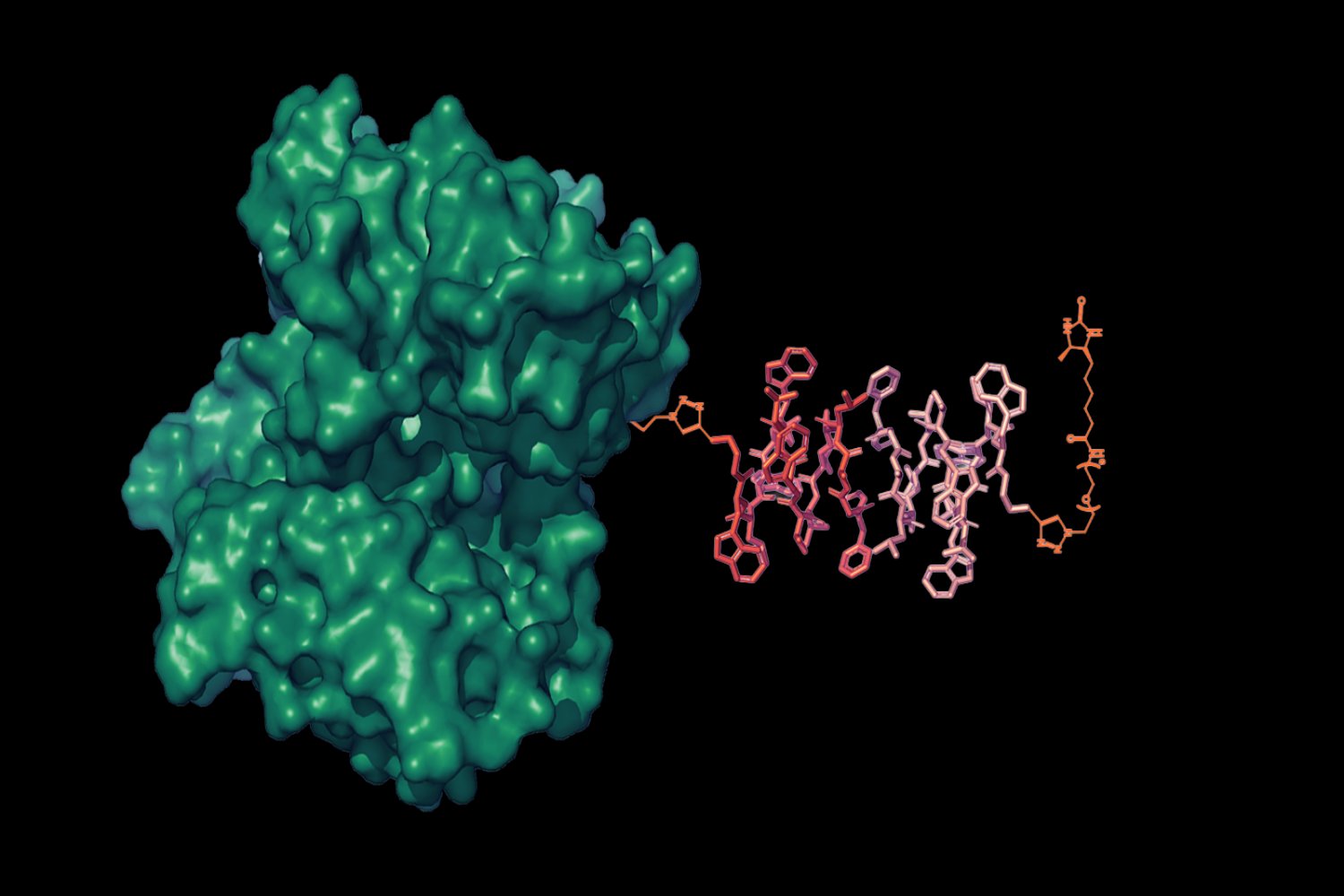
The new sensors, developed by postdoc Sayani Das and graduate student Jacob Cyert Simon, address this issue by allowing a single target molecule to affect multiple contrast agents. This is achieved by enclosing MRI contrast agents in liposomal nanoparticles filled with gadolinium, which enhances MRI signals from hydrogen atoms in water.
Das and Simon engineered water channels in these nanoparticles, which open or close based on the presence of a target molecule, affecting MRI signal brightness. They named these sensors liposomal nanoparticle reporters, or LisNRs, which react to specific molecules by altering water flow and thereby changing the MRI signal.
In experiments led by postdoc Miranda Dawson, the team used LisNRs to detect biotin in rats’ brains and bodies, showing the sensors’ amplifying capabilities. Jasanoff reports they achieved sensitivity about tenfold greater than conventional methods and believes further improvements are possible.
The sensors can be delivered throughout the body, making them useful for brain-wide and peripheral nervous system imaging. The next step involves creating LisNRs that respond to specific neurochemicals, starting with dopamine and glutamate, which are abundant and essential for neuron communication.
This research received funding from several MIT centers, including support for the involved postdoctoral fellows and graduate students.
Original Source: news.mit.edu