MIT researchers have developed a new technique called “implosion carving” that creates vacancies in materials, reducing them to approximately 1/2,000 of their original size. This advancement in nanotechnology could lead to devices for optical computing and visible light manipulation. The method utilizes photopatterning in a hydrogel, achieving features smaller than 100 nanometers, allowing the devices to bend light for optical computations.
Quansan Yang, a former MIT postdoc now at the University of Washington, emphasized the importance of creating nanostructures with resolutions under 100 nanometers for manipulating visible light. Researchers demonstrated a photonic device capable of a basic digit-classification task, with potential for future high-speed imaging and information processing applications. The study, co-authored by Gaojie Yang, was published in Nature Photonics.
Senior authors include Peter So from MIT’s Laser Biomedical Research Center and Edward Boyden, a Howard Hughes Medical Institute investigator. Photonic devices offer a promising, energy-efficient alternative to semiconductor chips, but current 3D manufacturing methods lack the required resolution for visible light manipulation. Existing techniques either lack 3D capabilities or don’t achieve necessary resolutions.
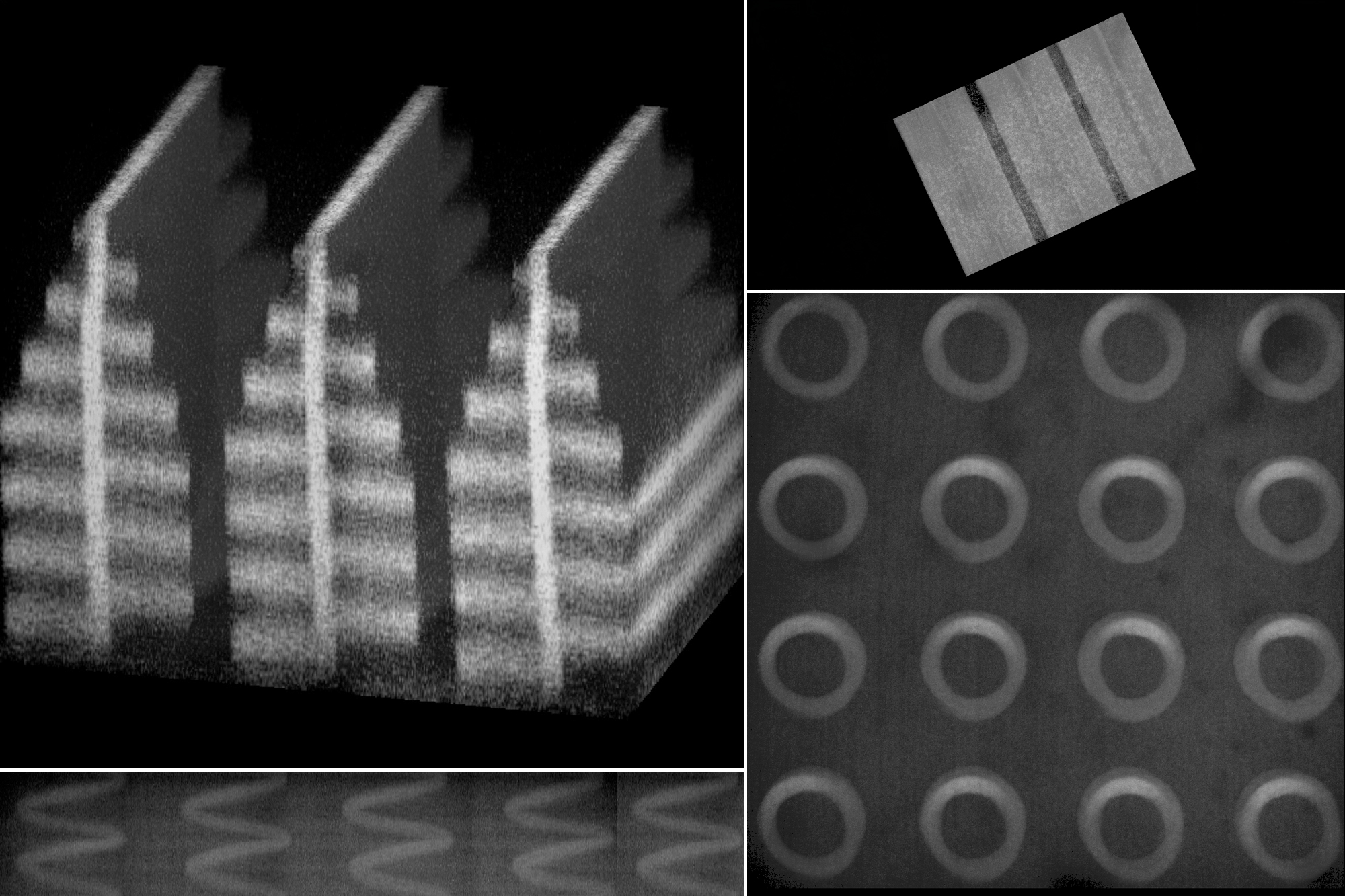
The team extended their 2018 “implosion fabrication” concept to create “implosion carving,” where lasers form vacancies in hydrogels. This process involves using a photosensitizing dye, a laser to excite the dye, and reactive oxygen species to break hydrogel bonds, forming vacancies. The gel is then shrunk through an ion solution and supercritical drying, achieving a significant volume reduction.
The researchers created various 3D shapes, including a helix and butterfly wing-inspired structure, demonstrating the technique’s flexibility. They also developed an optical device for digit classification, using patterned vacancies to diffract light and determine the output. This approach offers new design opportunities using deep-learning algorithms, says Dushan Wadduwage from Old Dominion University.
Future plans involve creating optical devices to classify cells in microfluidic devices, potentially identifying rare cells like circulating tumor cells. The method could also lead to high-throughput imaging for tissue analysis and, when adapted for hydrophobic polymers, create channels in 3D nanofluidic devices. The research was supported by various institutions, including the MIT-Fujikura Partnership Fund and the U.S. National Institutes of Health.
Original Source: news.mit.edu