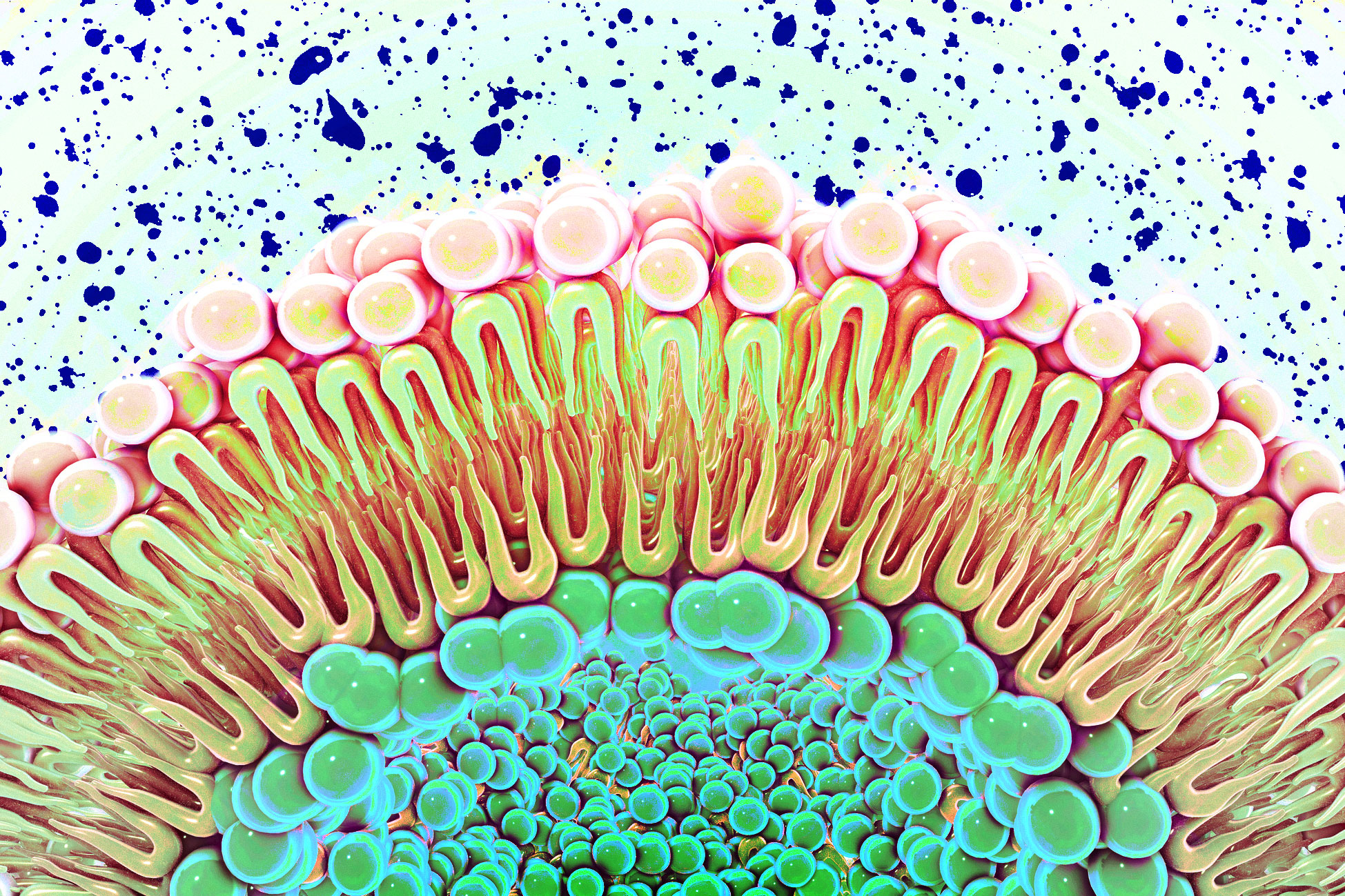
Cells are surrounded by a lipid membrane that provides both structure and a barrier between the cell and its surroundings. Recent evidence indicates these membranes also impact the behavior of protein receptors within them. An MIT study further supports this, revealing that altering the membrane’s composition can change the function of a receptor that encourages cell proliferation.
Researchers discovered that the epidermal growth factor receptor (EGFR) can become overactive when the cell membrane has an increased concentration of negatively charged lipids. This may explain why cancer cells with high levels of these lipids exhibit unchecked growth. “Traditionally, membranes were seen merely as scaffolds, but increasing observations suggest these lipids may influence receptor function,” says Gabriela Schlau-Cohen, senior author and MIT chemistry professor.
The study suggests potential tumor treatments by neutralizing negative charges to reduce EGFR signaling. Shwetha Srinivasan PhD ’22 led the study, published in eLife, with contributions from former MIT postdocs Xingcheng Lin and Raju Regmi, Xuyan Chen PhD ’25, and MIT associate professor Bin Zhang.
EGFR, present on cells lining body surfaces and organs, is one of several receptors that regulate cell growth. Certain cancers, like lung cancer and glioblastoma, overexpress this receptor, leading to uncontrolled cell growth. EGFR spans the cell membrane, making it difficult to study signaling across its entire structure.
Schlau-Cohen’s lab uses nanodiscs, self-assembling membrane models, to facilitate studying these processes. Embedding receptors in these discs allows for the examination of the full-length receptor. Single molecule FRET (fluorescence resonance energy transfer) is used to study receptor shape changes under different conditions by measuring energy transfer between fluorescent tags.
Previous research by Schlau-Cohen and Bin Zhang utilized single molecule FRET and simulations to demonstrate that EGFR binding to EGF induces a shape change in the receptor, activating cellular mechanisms that promote growth. The new study explores how altering membrane composition affects receptor function, focusing on negatively charged lipids.
Normally, membranes comprise about 15 percent negatively charged lipids. The researchers found that increasing this to 60 percent locked the EGFR receptor in an active state, constantly signaling growth even without EGF binding. Many cancer cells have elevated levels of these lipids, potentially explaining why they grow uncontrollably. “With high levels of negatively charged lipids, the receptor remains in an open, growth-signaling conformation regardless of ligand binding,” Schlau-Cohen explains.
The study also investigated cholesterol’s role in EGFR function. Elevated cholesterol levels in nanodiscs increased membrane rigidity, which suppressed EGFR signaling. The research received funding from the National Institutes of Health and MIT’s Department of Chemistry.
Original Source: news.mit.edu