



Researchers in tissue engineering aim to create living organs and tissues from cells to replace damaged body parts. While artificial livers, kidneys, and muscles have been successfully grown, replicating complex blood vessel networks has been elusive. Without these networks, artificial tissues can’t function properly. MIT engineers have discovered a method to control blood vessel growth by mechanically stretching them.
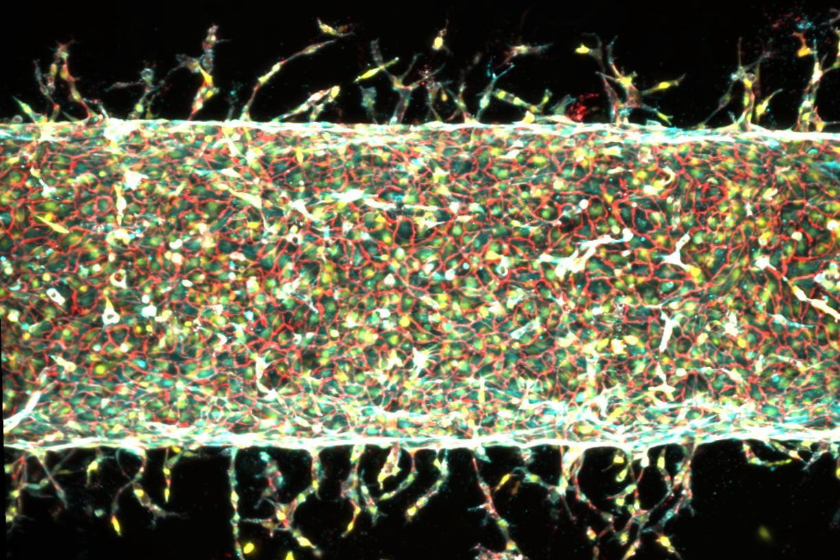
The team developed a “blood vessel on a chip,” featuring a central artery made from human endothelial cells within a gel containing a small magnet. By moving the gel with an external magnet, they observed that the artery sprouted smaller capillaries. Adjusting the direction and degree of stretching allowed them to influence the growth and pattern of these vessels. Their findings, published in the Proceedings of the National Academy of Sciences, provide a new approach to designing artificial blood vessels.
According to Ritu Raman, associate professor of mechanical engineering at MIT and co-lead author, organized blood vessel networks are essential for healthy tissues, but current methods don’t support their fabrication in engineered tissues. The ability to guide blood vessel growth using physical cues could lead to scalable and reproducible engineered tissues for medical implantation.
Conventional techniques struggle to grow and control fine blood vessel networks. Although 3D printing can create larger vessels, it lacks the precision for tiny capillaries. While some progress has been made growing vessels from cells in nutrient-rich environments, precise control remains challenging. Raman’s team wondered if mechanical stimulation, used in previous projects on artificial muscles, could also manipulate blood vessel growth.
Using a similar gel setup with embedded magnets, researchers grew a central artery on a chip and observed new vessel sprouting under mechanical manipulation. Stretching the artery enhanced capillary growth, with variations in stretching affecting vessel number and direction. The study suggests that mechanical forces are crucial in directing blood vessel growth.
Further investigation focused on the PIEZO1 gene, linked to a cell’s response to mechanical pressure. After consulting Nobel laureate Ardem Patapoutian, the team explored the gene’s role in blood vessel growth. By reducing PIEZO1 activity, they confirmed fewer vessels grew under mechanical stimulation, indicating its involvement in vessel development.
With this new ability to control blood vessel growth, the researchers plan to extend their method to support artificial organ and tissue development. Co-author Jessica Shah notes that they are now exploring how precisely patterning blood vessel growth can enhance muscle function. The research received support from the U.S. Department of War Army Research Office Early Career Program and PECASE Grant, and a Department of War DURIP Program Grant.
Original Source: news.mit.edu