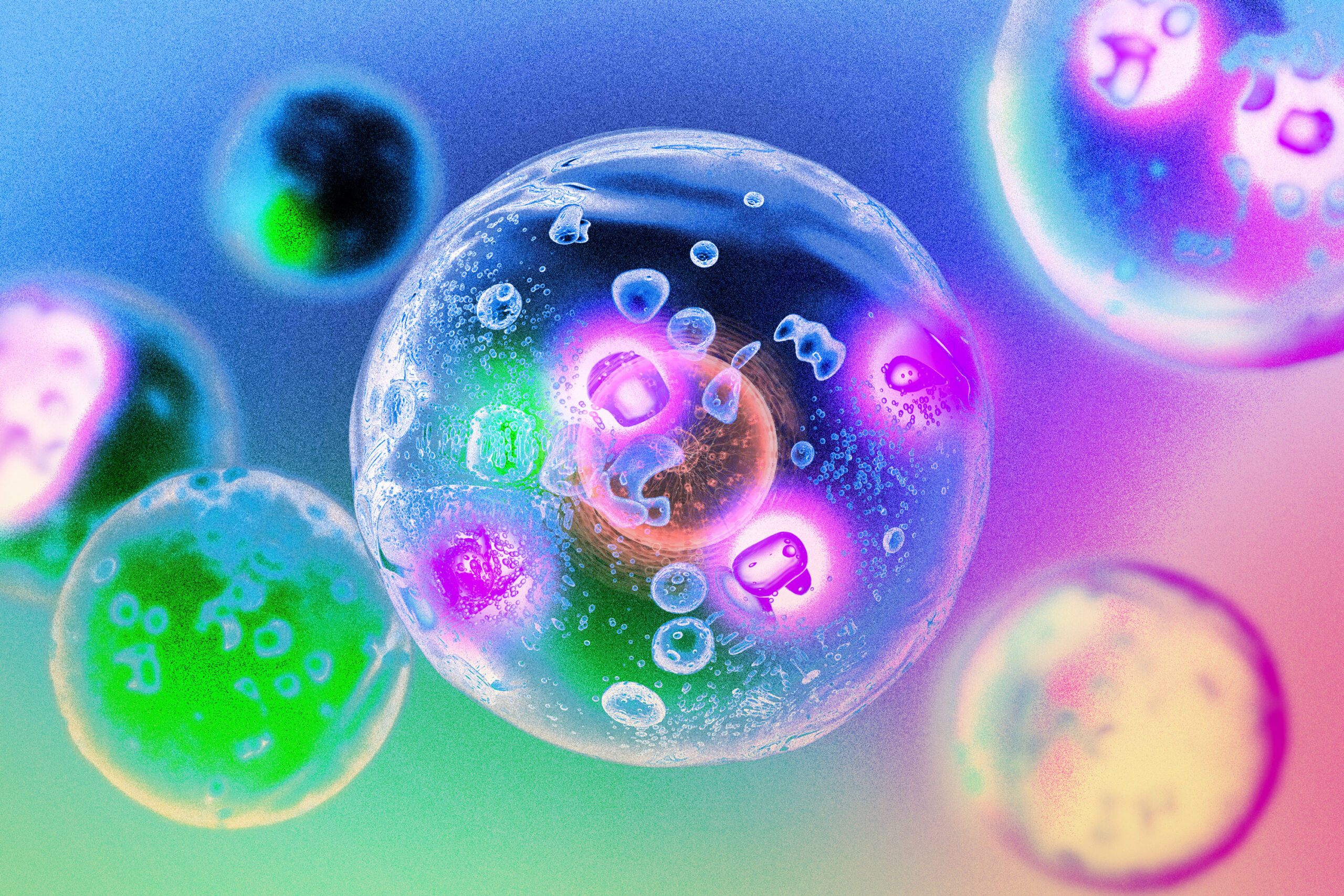
In recent years, scientists have uncovered that cells maintain order through a process called phase separation. This is akin to oil droplets forming in vinegar, where proteins within cells create concentrated droplets for organization. A study by MIT researchers has revealed that this droplet formation is crucial for the function of enzymes known as kinases. By condensing into droplets, kinases can optimize conditions to catalyze reactions faster, enhancing cell signaling pathways. Sometimes, this formation alters the reactions kinases perform.
“Many biological molecules have this propensity to spontaneously separate. We were really interested in asking, if we have these kinases forming droplets, what is the consequence of that in the context of signaling?” says Lindsay Case, an assistant professor at MIT and senior author of the study. Understanding droplet formation could aid in designing kinase-targeting drugs, as some kinases become overactive in cancer cells. “Understanding the chemistry of these compartments, and what molecules go into them and what molecules don’t go into them, could help us design drugs that better localize to their target of interest,” Case adds. Nicholas Lea, an MIT graduate student, led the study published in Cell Reports.
Since her graduate studies, Case has explored how molecular organization within cells affects their function. During her postdoctoral research, she examined phase separation’s impact on a signaling pathway that enables cells to sense their attachment to the environment. Proteins involved in this pathway include kinases, which activate other proteins by adding phosphate groups. Kinases can self-activate through autophosphorylation.
“Inside of the cell, you have these kinase molecules that are responsible for carrying a signal through the cell, and we know that the organization of these molecules changes. When the information is present, they’re organized in a different way than when the information is not present,” Case explains. “We think that having the right molecules in the right place is incredibly important for the right biochemistry to occur.” Phase separation is a method cells use for organization, similar to oil droplets forming in salad dressing to avoid water-based vinegar. Proteins can phase separate at high concentrations, leading them to form dense droplets in the cytoplasm.
Case theorized that phase separation, which clusters kinases densely, might enhance enzyme activity by increasing the likelihood of their interaction and phosphorylation. Case and Lea tested this with focal adhesion kinase (FAK), which activates when cells attach to their environment, prompting pro-growth and pro-survival signals. In cancer cells, this pathway can malfunction, enabling cells to proliferate after detachment.
Scientists knew that proper cell attachment causes FAK accumulation at the membrane. The MIT team replicated this by overexpressing FAK in unattached cells, causing the kinase to phase separate into droplets, activating the pro-growth signal. “It was surprising that just by condensing this protein into a droplet, you can actually turn on a signaling pathway that should be turned off,” Case says. If FAK concentration is too high, droplets persistently form, continuously signaling regardless of receptor control.
The findings imply that FAK overexpression in cancer cells may lead to phase separation, promoting cancer progression and metastasis. “It may be that for some kinases, you’re not supposed to form these droplets in the cytoplasm because it leads to this always-on signal, and then the cells no longer listen to the information coming from the environment,” Case notes. Disrupting FAK’s droplet-forming ability could offer a new cancer drug development strategy.
The researchers also examined two other kinases, Mst2 and Abl, finding that they too could phase separate at high concentrations, enhancing their activity. While FAK’s cytoplasmic phase separation may be cancer-specific, Mst2 utilizes this strategy in healthy cells to regulate the Hippo signaling pathway, which supports cell growth and survival.
For both Mst2 and Abl, phase separation allows the enzymes to phosphorylate additional targets, potentially activating different signaling pathways. “It’s not just that you’re getting faster phosphorylation, but in those cases, the patterns of what is actually getting phosphorylated were very different inside of the droplet compared to what might be happening in a non-droplet context,” Case says. “The kinase is able to phosphorylate amino acid residues beyond the set of canonical sites that have been described before.”
The study also found that droplet formation attracts high ATP concentrations, as kinases contain floppy sections with positively charged amino acids that draw in negatively charged ATP. Using a machine-learning model, researchers predicted that approximately 45 percent of the 500 human cell kinases could form similar droplets, aided by their positive charge, which might recruit ATP.
Case aims to explore designing drugs that mimic ATP’s droplet-attracting ability, potentially reducing drug side effects. “By localizing drugs to the compartment where your target localizes, that could reduce off-target effects by concentrating the drug with the target of interest and reducing interactions with other molecules,” Case states. The research received funding from several sources, including the Searle Scholars Program Award and the U.S. Air Force Office of Scientific Research.
Original Source: news.mit.edu