



Approximately 85,000 individuals in the United States receive a bladder cancer diagnosis annually. Although treatments are frequently effective, bladder cancer has a recurrence rate of about 50% within five years post-treatment, ranking it among the most recurrent cancers. This recurrence contributes to its high treatment costs. Researchers at MIT have developed a novel method for monitoring patients, potentially allowing for earlier detection of returning tumors. The technique involves a catheter with nanosensors capable of identifying low concentrations of a protein linked to bladder cancer and visualizing their tissue location.
The MIT team asserts that their sensing method is almost 50,000 times more sensitive than traditional urinalysis, which is commonly used for monitoring bladder cancer. In animal tests, the fluorescent signals from the sensors provided precise tumor location information within the bladder lining, creating a chemical map. “It’s like a camera for molecules instead of light,” remarked Michael Strano, the Carbon P. Dubbs Professor of Chemical Engineering at MIT. Strano led the study, published in Nature Nanotechnology, alongside lead authors Wonjun Yim and Hohyung Kang, with contributions from other MIT researchers.
This new detection technique employs carbon nanotubes, which are tiny cylinders made of carbon that emit fluorescence when exposed to laser light. Over the past decade, Strano’s lab has adapted these nanotubes to detect various molecules by coating them with “synthetic antibodies” that bind to specific targets. For this study, the team developed a sensor to detect nuclear matrix protein 22 (NMP-22), an FDA-approved bladder cancer biomarker. NMP-22 is often diluted in urine, which complicates tumor detection until advanced stages.
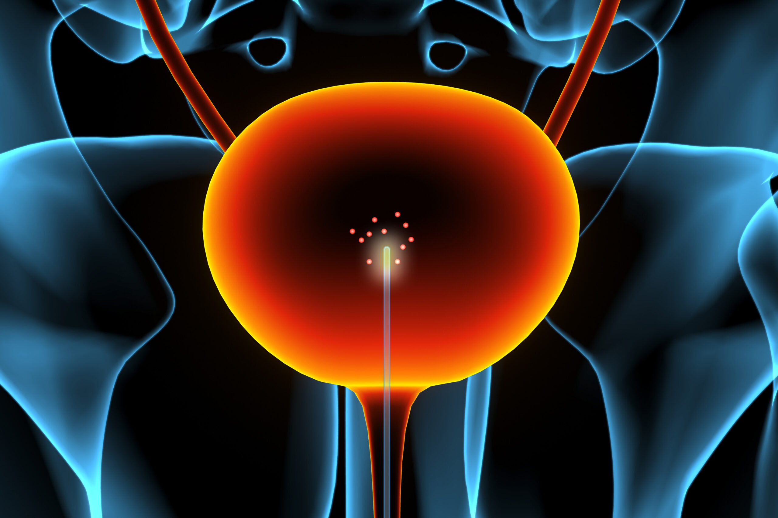
To detect tumors earlier, the MIT team created a device to deploy sensors inside the bladder, allowing them to detect NMP-22 at elevated concentrations near tumors. Their device uses a urinary catheter coated with nanotubes and a small rotating ball lens to emit and absorb fluorescent light from the nanosensors, enabling the mapping of biomarker locations. These chemical images reveal both the presence and location of cancerous cells. Strano explained that detecting chemical signals before a tumor becomes visible allows for precise location mapping.
Animal tests demonstrated that this detection method is 180 times more sensitive than standard urinalysis by detecting biomarkers directly in the bladder. This sensitivity could identify tumors as small as 16 square millimeters. Strano’s lab is now working on a more compact imaging system for easier use in clinical settings and aims to integrate the sensors into cystoscopes, which are used to visualize bladder tumors. This advancement could enable earlier detection and treatment of recurring tumors, reducing monitoring costs.
Strano highlighted the potential for faster, less invasive, and more cost-effective screening in doctor’s offices. Daniel Heller from Weill Cornell Medicine, not involved in the research, noted that bringing the sensor to the patient enhances diagnostic effectiveness, potentially improving cancer treatment. The approach could also be adapted to detect other diseases, such as cardiovascular or gastrointestinal conditions, by modifying the nanosensors on the catheter. “The beauty of polymer chemistry is that if we understand the molecular structures of target biomarkers and the design principles of binding sites, we can develop new sensors tailored to different diseases,” remarked Yim.
Original Source: news.mit.edu