Relaxor ferroelectrics have long been integral to technologies such as ultrasounds, microphones, and sonar systems due to their distinct atomic structures. Until now, direct measurement of this structure has been elusive. A team from MIT and other institutions has successfully mapped the three-dimensional atomic structure of a relaxor ferroelectric for the first time. Their findings, published in Science, offer a foundation for improving models used in creating future computing, energy, and sensing technologies.
James LeBeau, MIT’s Kyocera Professor of Materials Science and Engineering, remarked, “Now that we have a better understanding of exactly what’s going on, we can better predict and engineer the properties we want materials to achieve.” The research community is still developing ways to engineer these materials, but accurate models are crucial for predicting their properties, LeBeau emphasized.
The researchers employed a new technique to explore the distribution of electric charges within the material, uncovering unexpected chemical disorder. Michael Xu PhD ’25 and Menglin Zhu, both MIT postdocs and co-first authors, noted that merging experimental data with simulations allowed them to refine models for better predictive accuracy.
Other contributors to the study include MIT students Colin Gilgenbach and Bridget R. Denzer, Yubo Qi from the University of Alabama at Birmingham, Jieun Kim from the Korea Advanced Institute of Science and Technology, Jiahao Zhang, formerly of the University of Pennsylvania, Lane W. Martin from Rice University, and Andrew M. Rappe from the University of Pennsylvania.
Simulations of relaxor ferroelectrics show that the interactions of charged atoms in nanoregions enhance energy storage and sensing abilities when an electric field is applied. These nanoregions’ specifics have been challenging to measure directly until now.
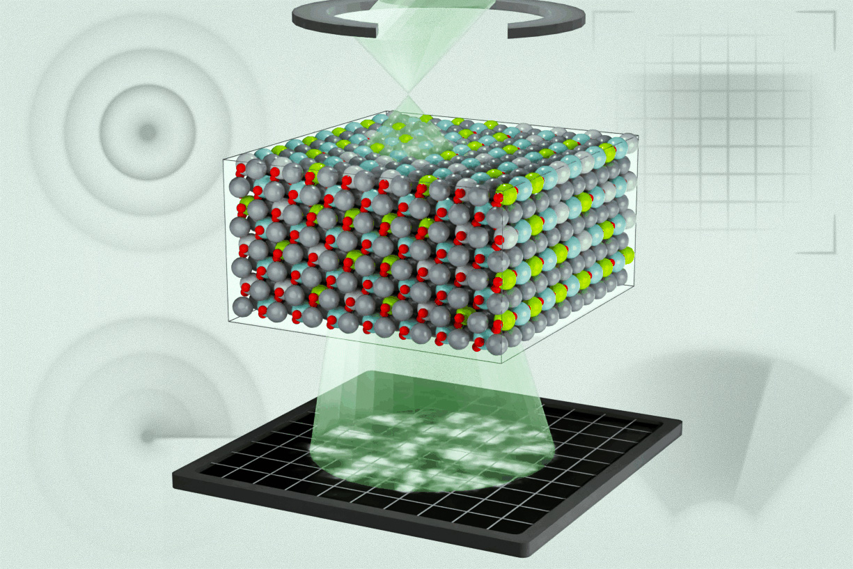
In their Science publication, the researchers examined a lead magnesium niobate-lead titanate alloy, a relaxor ferroelectric material used in various applications. They utilized multi-slice electron ptychography (MEP), a technique that involves moving a nanoscale high-energy electron probe over the material and capturing the resulting diffraction patterns.
Zhu explained, “We do this in a sequential way, and at each position, we acquire a diffraction pattern.” This process creates overlapping regions containing enough information to reconstruct three-dimensional data about the material and the electron wave function.
The study revealed a hierarchy of chemical and polar structures from atomic to mesoscopic scales. The team found that many regions of differing polarization were smaller than previously predicted, and they used this new data to refine simulation models under various conditions.
Xu noted that earlier models included random polarization regions without correlations. Now, they can provide detailed information on how chemical species affect polarization based on atomic charge states.
Zhu highlighted the potential of electron ptychography for studying complex materials, suggesting it could lead to new research directions in disordered materials. Xu added, “This study is the first time in the electron microscope that we’ve been able to directly connect the three-dimensional polar structure of relaxor ferroelectrics with molecular dynamics calculations.”
The researchers believe this approach could eventually aid in engineering materials with advanced electronic properties for enhanced memory storage, sensing, and energy technologies. LeBeau pointed out that with the increasing complexity in material design, accurate models are essential for understanding material behavior and validating designs.
The project received support from the U.S. Army Research Laboratory, the U.S. Office of Naval Research, the U.S. Department of War, and a National Science Graduate Fellowship, with resources provided by MIT.nano facilities.
Original Source: news.mit.edu