In the primary visual cortex, known for its role in processing visual information, not all neurons engage in processing what the eyes see. This may be due to the vast array of inputs each neuron receives through thousands of synapses, forcing them to choose between processing visual and other types of information. A recent study conducted on mice by neuroscientists at MIT’s Picower Institute for Learning and Memory demonstrates how neurons organize these inputs to function effectively.
Neuroscientists are eager to understand what prompts neurons to partake in the brain’s computational functions, says senior author Mriganka Sur, a professor at MIT’s Department of Brain and Cognitive Sciences. Neurons contribute to brain circuits by generating an electrical action potential. Sur explains that the structure and organization of neuron inputs are crucial for brain circuits to process information. The study, published in iScience, offers insights into this process.
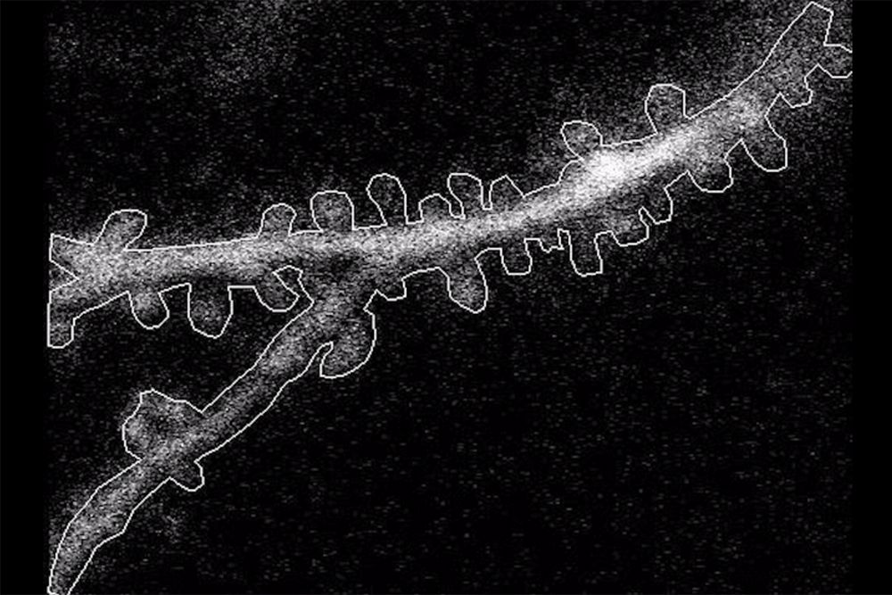
Led by postdoc Kyle Jenks, the research team used imaging techniques to observe how neurons’ cell bodies and individual synapses on dendritic spines reacted when mice viewed moving visuals. They studied both visually responsive neurons and those that were unresponsive but had visually responsive spines. This allowed for an analysis of factors influencing synapse formation and its impact on cell body responses.
Jenks and his team engineered neurons so their dendritic spines would glow in response to calcium activity, indicating synapse activity. This was done for both the spines and the cell body, allowing the team to monitor responses to black and white grating patterns viewed by the mice. The study tracked 11 neurons active to visual input and 11 that were not, leading to several findings.
The proximity to the soma was significant: spines closer to the soma were more likely to correlate with its activity. Additionally, neurons that responded to visual input had spines forming clustered groups of correlated responses within 5 microns. Neurons have two types of dendrites: apical and basal. Basal dendrites, receiving more raw visual input, differed from apical dendrites, which had more visually responsive spines in responsive neurons. However, both dendrite types followed the distance rule from the soma.
Orientation selectivity emerged as the most crucial factor in determining the correlation between a spine’s responsiveness and the soma. The research team concluded that synaptic inputs to layer 2/3 neurons in the mouse visual cortex are organized based on factors like somatic responsiveness and stimulus selectivity. These findings could aid in studying visual processes and genetic mutations affecting neuron connections.
The study was supported by the National Institutes of Health, the Simons Foundation Autism Research Initiative, and the Freedom Together Foundation. In addition to Sur and Jenks, authors include Gregg Heller, Katya Tsimring, Kendyll Martin, Asrah Rizvi, and Jacque Pak Kan Ip. Sur notes that these rules can serve as benchmarks for future research on the effects of genetic mutations on vision.
Original Source: news.mit.edu