People at high risk for breast cancer might find that annual mammograms are insufficient for early tumor detection. An MIT team has developed portable ultrasound detectors to facilitate more frequent imaging. Their recent paper highlights improvements in image resolution, aiding in the identification of tumors, cysts, and microcalcifications. The team also designed a user-friendly interface for the ultrasound probe, allowing even those without ultrasonography expertise to operate it. This system could promote earlier detection and enable ongoing monitoring after breast cancer treatment, whether in a clinical setting or at home.
“At each time interval, the computer interface guides you to position the device in exactly the same location, which is important for the longitudinal monitoring of a given tissue. It’s very intuitive and quite easy to use,” explained Canan Dagdeviren, an MIT associate professor and the study’s senior author. The lead authors, former MIT postdoc Md Osman Goni Nayeem and MIT graduate students Shrihari Viswanath and Hyeokjun Yoon, published their findings in Nature Communications.
While many undergo annual mammograms to screen for breast cancer, some cancers emerge between these screenings, known as interval cancers, which are often more aggressive and represent 20 to 30 percent of all cases. After losing an aunt to such a cancer in 2015, Dagdeviren sought a better screening method for women with dense breast tissue that could be used more frequently than mammography. She opted for ultrasound, which creates tissue images using sound waves. Traditionally, ultrasounds require large equipment and skilled operators.
Earlier this year, Dagdeviren’s lab showcased a compact ultrasound probe linked to a module slightly bigger than a smartphone, capable of generating 3D breast images by scanning just a few locations. Their latest study in Nature Communications reports advancements leading to higher resolution images and easier usability.
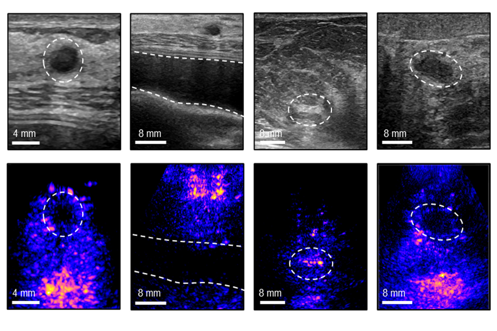
A significant improvement was adding a “backing layer” to the ultrasound transducer, which helps focus sound waves, enhancing image quality and resolution. It also broadens the range of frequencies absorbed and minimizes acoustical and electrical noise. “With the backing layer, the device produces more accurate and sharper images, with a wider operating range of frequencies,” said Nayeem.
To further refine image quality, the researchers developed a beamforming algorithm that adjusts for varying sound wave speeds through different tissues. “We aim to predict the speed of sound properties of the tissue you’re imaging, and use that to reconstruct the image more accurately. We see up to a 10 percent improvement in the resolution by applying this technique,” Viswanath noted.
The team tested the system with 10 volunteers who were unfamiliar with ultrasound technology, asking them to find micro targets in a “tissue phantom” designed to mimic human tissue. The volunteers had a higher success rate using the new system compared to a standard ultrasound probe.
The updated system includes a computer interface that guides users to correctly position the probe, crucial for monitoring treatment progress or known abnormalities. In a trial with seven participants, users accurately placed the probe each time. “Conventionally, you need an operator to move the probe around the breast, but we made a computer-vision interface for users to do it by themselves. This is very user-friendly and it shows live images on the screen,” Yoon explained.
Future versions might feature an interface compatible with cellphones or tablets, enhancing portability and access in regions lacking trained ultrasound technicians. Dagdeviren and her students plan to establish a company to commercialize this technology, initially targeting breast cancer diagnosis with potential expansion to other applications.
“The technology is so versatile that it can be used for any soft tissue imaging, from ovarian cancer to measuring endometriosis progression, or fetal monitoring,” Dagdeviren stated. The research received funding from the National Science Foundation, the 3M Non-Tenured Faculty Award, the Lyda Hill Foundation, the MIT Media Lab Consortium, and a Tata Center Technology and Design Fellowship.
Original Source: news.mit.edu