MIT researchers have uncovered an unexpected phenomenon in optical physics that may lead to a new, faster, and higher-resolution bioimaging method. They found that, under specific conditions, chaotic laser light can spontaneously organize into a focused “pencil beam.” This beam allowed them to capture 3D images of the human blood-brain barrier 25 times faster than the standard method, with similar resolution.
By enabling real-time visualization of individual cells absorbing drugs, this technology could help in verifying whether new treatments for neurodegenerative diseases like Alzheimer’s and ALS effectively reach their brain targets. “The common belief is that increasing laser power leads to chaotic light, but we showed otherwise. We embraced the uncertainty and let the light organize itself for a new bioimaging solution,” says Sixian You, assistant professor at MIT and senior author of the study published in Nature Methods.
The research team included lead author Honghao Cao and several other MIT affiliates, along with Subhash Kulkarni from Harvard University and Roger Kamm from MIT. The discovery began with a surprising observation when Cao pushed a multimode fiber to its power limits, expecting disorder, but instead, the light formed a sharp beam.
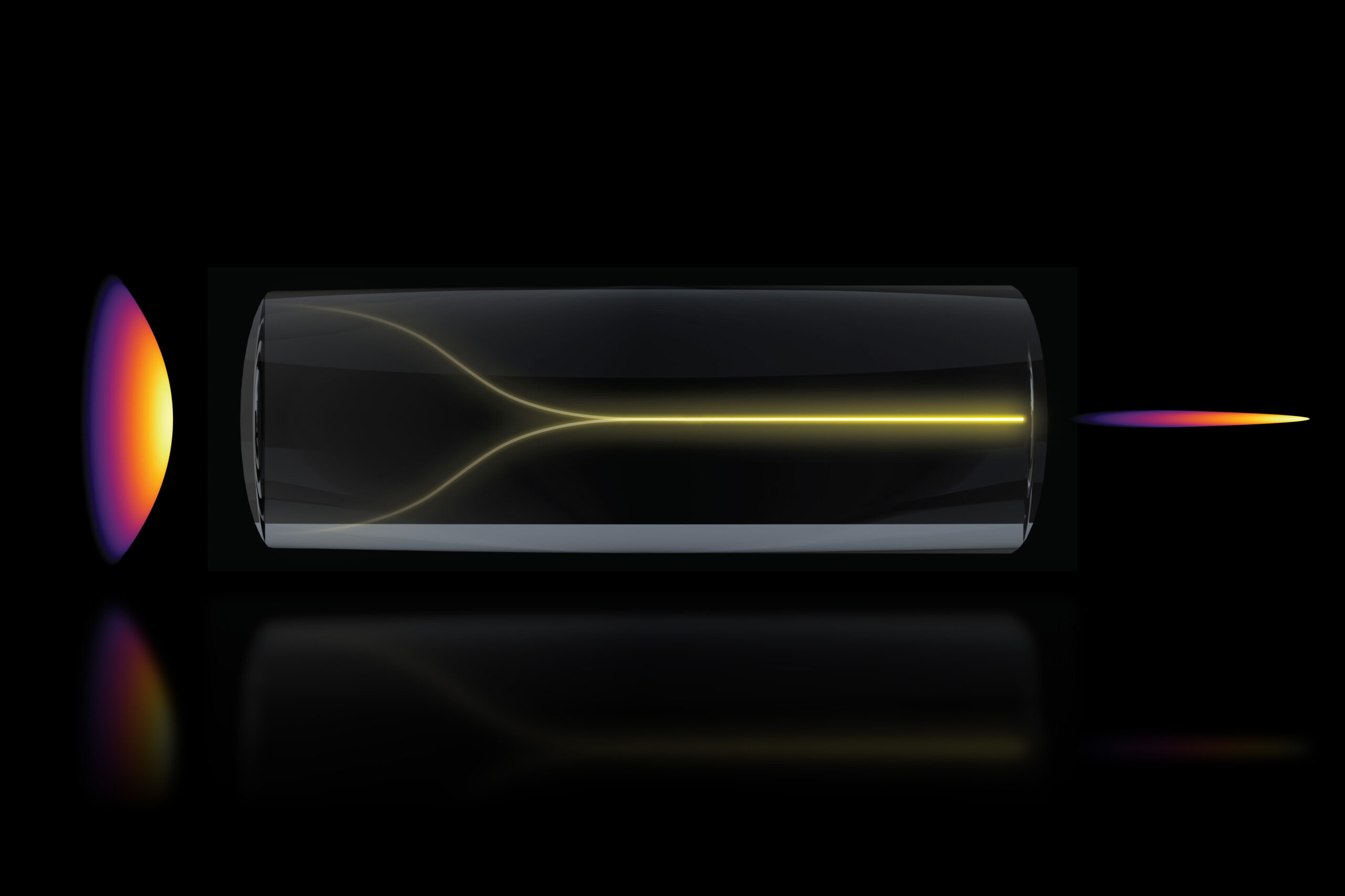
Typically, more power results in a scattered beam due to fiber imperfections. However, Cao found that by increasing the power near the fiber’s burning point, the light formed a needle-like beam. “Disorder is intrinsic to these fibers, but self-organization provides a stable, ultrafast pencil beam without complex components,” You explains.
Two precise conditions were needed to replicate this phenomenon: the laser must enter the fiber at a perfect angle and the power must be increased to interact with the fiber glass. “At critical power, nonlinearity counters intrinsic disorder, transforming the input beam into a self-organized pencil beam,” says Cao.
Usually, experiments at lower power levels don’t reveal self-organization, and precise alignment isn’t common due to the fiber’s power capacity. However, these methods together create a stable pencil beam without intricate light engineering. “The charm of this method is its simplicity, achievable with a standard optical setup,” You says.
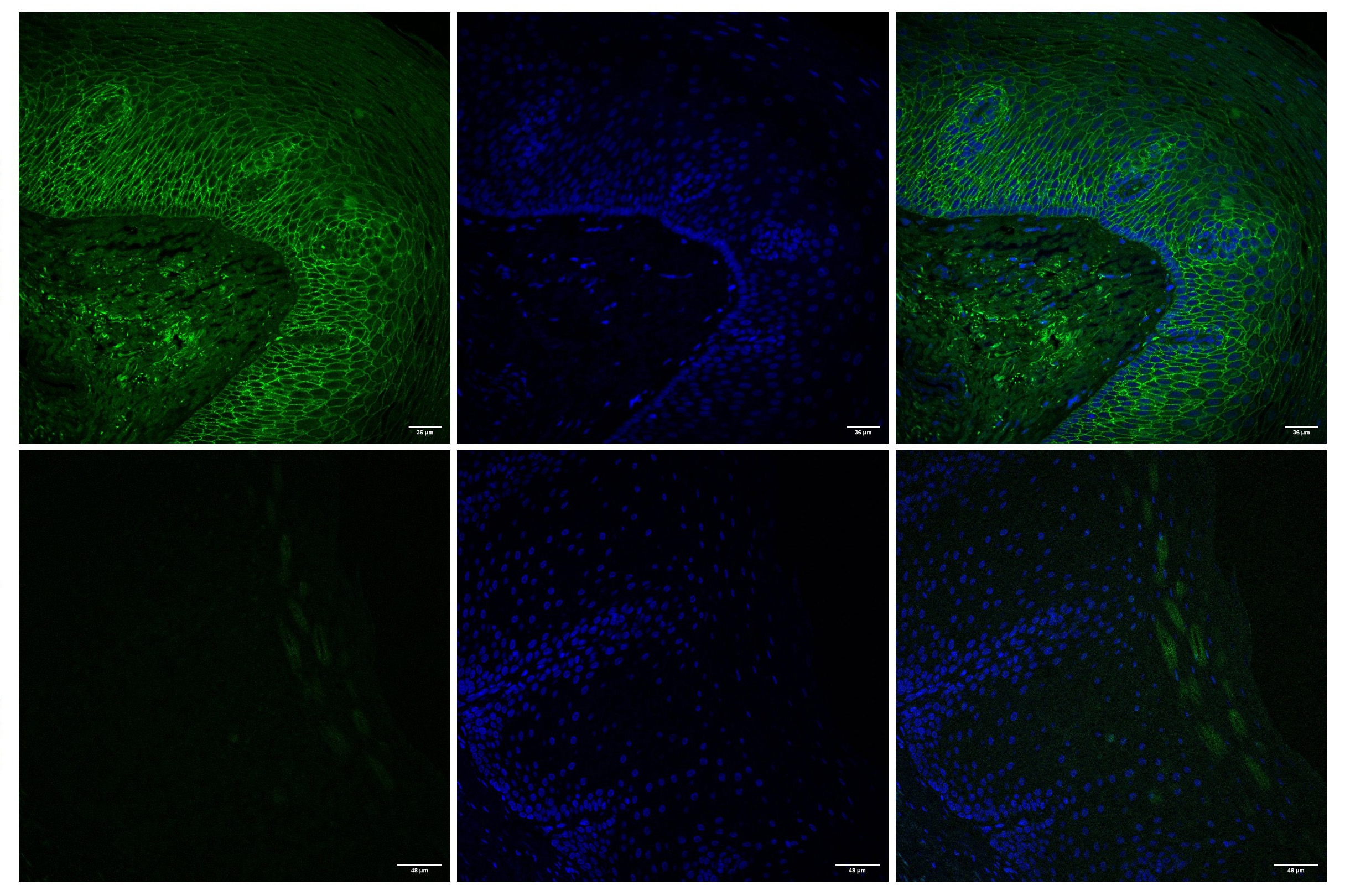
Characterization experiments showed the pencil beam was more stable and high-resolution, with less “sidelobe” distortion. The researchers used it for biomedical imaging of the blood-brain barrier, a protective cell layer blocking toxins and many medicines. Standard settings capture only a 2D vasculature section at a time, You explains.
The new technique allowed ultrafast, high-precision tracking of cell protein absorption in real-time. “The pharmaceutical industry is keen on using human models for drug screening, as animal models often fail in humans. This method, not requiring fluorescent tags, is revolutionary,” says Kamm.
“This approach is not just for the blood-brain barrier; it tracks diverse compounds and targets in engineered tissues, enhancing biological engineering,” adds Spitz. The team produced cellular-level 3D images faster and with higher quality than other methods. “Our method overcomes the tradeoff between resolution and focus depth,” You says.
Looking ahead, the researchers aim to understand the pencil beam’s fundamental physics and self-organization mechanisms. They also plan to explore its use in other applications, like imaging brain neurons, and work on commercializing the technology. “You’s group demonstrated the beam’s value for microscopy reliant on light intensity, showing advantages over ordinary lasers. Understanding these new beams will be scientifically intriguing,” says Frank Wise, a professor at Cornell University, not involved in the study.
Original Source: news.mit.edu