








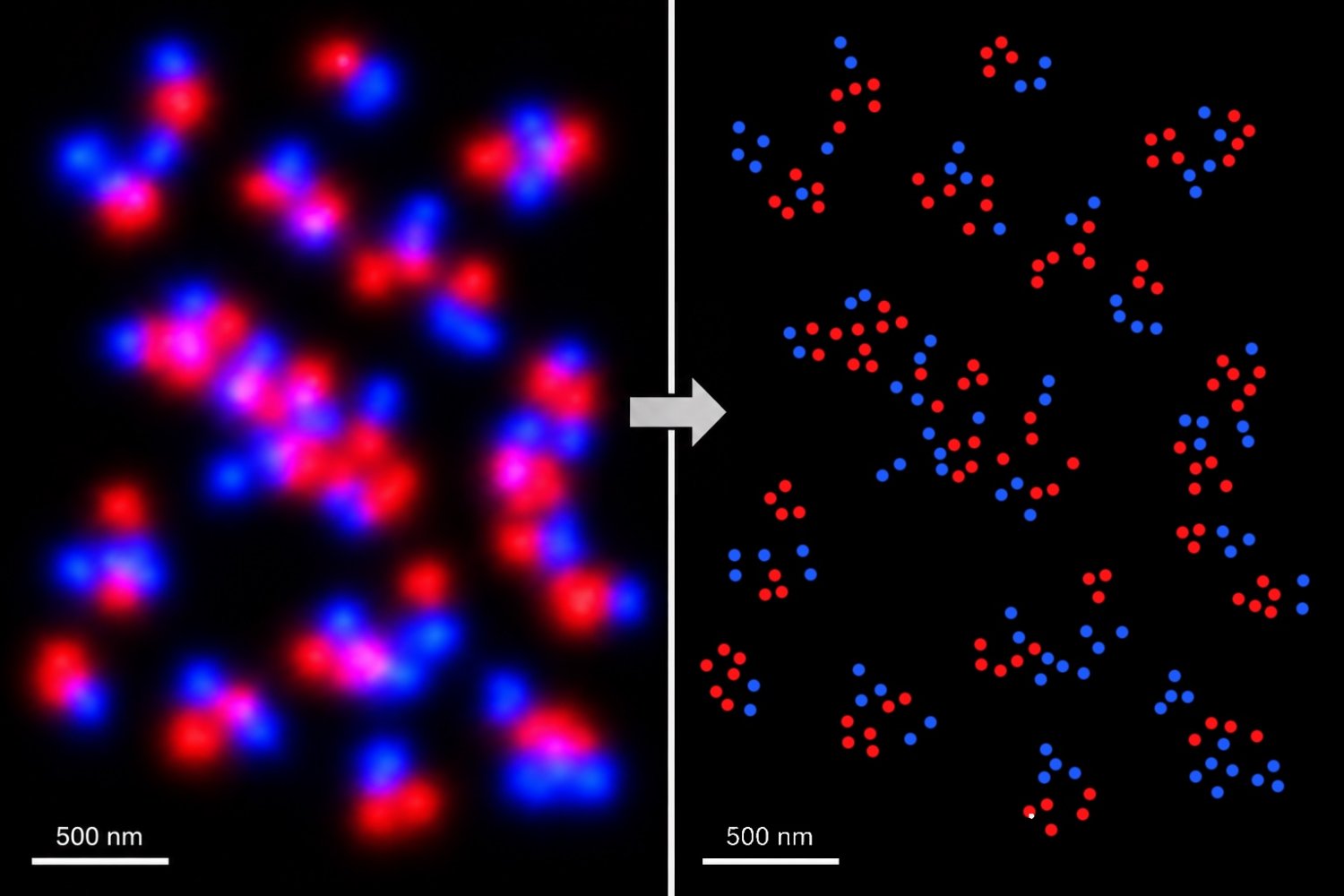
Researchers from the lab of Sam Peng, who holds the Pfizer Inc. – Gerald Laubach Career Development Assistant Professorship in Chemistry at MIT and is a key member of the Broad Institute of MIT and Harvard, have invented a revolutionary super-resolution imaging method. This technology allows for the visualization of molecular structures with sub-angstrom precision, surpassing the typical nanometer limits of standard fluorescent dyes by three magnitudes, while significantly simplifying the imaging process.
Unlike conventional dyes that quickly degrade under light, limiting data collection, the U-STORM (Upconversion enabled Stochastic Optical Reconstruction Microscopy) platform uses a new kind of engineered upconverting nanoparticles (UCNPs) that spontaneously and indefinitely blink. This advancement marks a significant change in both optical materials and biological imaging. Details of the study appeared in Nature Nanotechnology on July 27.
For many years, scientists believed that upconverting nanoparticles were entirely photostable and non-blinking, making them unsuitable for localization-based super-resolution microscopy methods like STORM. These techniques depend on the random “blinking” of light emitters to differentiate closely-packed molecules. “Our lab has been focused on overcoming these challenges,” Peng states. “We questioned whether it was possible to create a super-resolution imaging platform that is long-lasting, multicolor, simple to use, and achieves very high localization precision without additional imaging buffers or optical controls.”
By carefully controlling the composition of nanoparticles, the MIT and Broad Institute team found that small (~10nm) core-shell particles can be made to blink spontaneously under continuous near-infrared light. This blinking persists indefinitely, eliminating the need for complex imaging buffers, oxygen scavengers, or external optical modulation.
An angstrom, a small unit used in chemistry to measure atomic sizes and distances, is now achievable with U-STORM’s indefinite blinking, allowing researchers to gather over 88,000 localization events from the same particle, achieving an unprecedented localization precision of 0.6 Å. Unlike traditional multicolor super-resolution imaging, which requires various costly lasers and precise optical alignment, U-STORM operates using a single near-infrared laser, simultaneously exciting nanoparticles that emit different colors, thus reducing the experiment’s complexity.
Instead of capturing images sequentially in multiple rounds for multicolor imaging, U-STORM captures multiple colors at once. The researchers demonstrated this by mapping epidermal growth factor receptor dimers and multimers in biological samples under physiological conditions without needing specialized imaging buffers.
This research not only pushes the limits of microscopy but also introduces a new design approach for lanthanide nanomaterials. The team is working to expand the color range, make the particles smaller and brighter, and use U-STORM to explore complex nanoscale protein structures and cellular signaling pathways. U-STORM offers laboratories around the world a straightforward yet powerful tool for high-precision molecular imaging.
Original Source: news.mit.edu